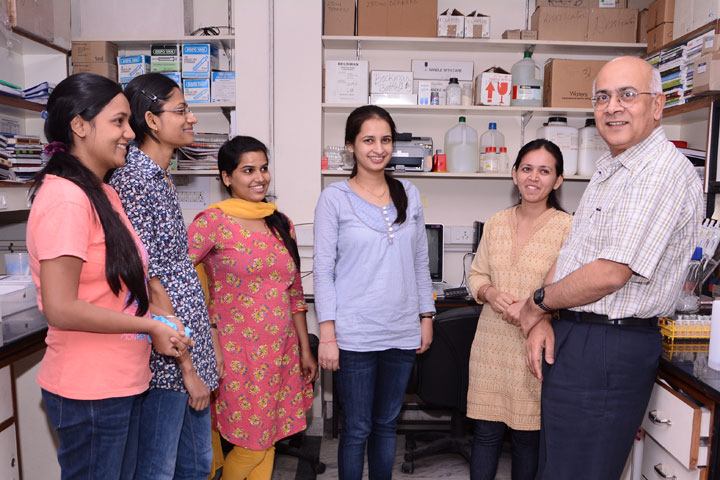
Associate Professor
E-mail: prem dot kaushal at rcb dot res dot in
Website: http://the-kaushal-lab.co.in/
Our research focuses on protein synthesis and ribosome assembly in pathogenic microbes, mainly Mycobacterium tuberculosis (Mtb), which causes the deadliest disease, tuberculosis, and Entamoeba histolytic (Eth), which causes amoebiasis. Protein synthesis is a fundamental cellular process that consumes nearly half of the cell's resources, and ~ 40% of the known antibiotics target the protein synthesis machinery, the ribosome. The research aims to find how pathogens survive under stresses by regulating translation machinery and exploit these features for novel inhibitor design.
In Mtb, the research focuses on understanding how the pathogen survives for decades in the hostile environment of the host macrophage in a dormant state. It is also believed that nearly one-third of the world’s population possesses the dormant Mtb, which serves as a huge reservoir for tuberculosis infection. We have reported a cryo-EM structure M. smegmatis ribosome, in complex with RafH protein at 2.8 Å resolution in Nature Communication . RafH is a hypoxia stress induced ribosome hibernation promotion factor. In the near future, we would like to understand mycobacterial survival under nutrition starvation and multiple stresses.
In Eth, our laboratory has reported the cryo- EM structure of its ribosome at 2.8 Å resolution, preprint available in bioRxive. This study reveals several unique features such as rRNA triple helix and helical assembly of Eth ribosome. In future we would like to determine its functional complexes, and understand the structural based of helical assembly.
Ribosomes, the mega Dalton sizes complexes, must assemble accurately to perform error-free translation. Defects in ribosome assembly, are associated with diseases such as cancer, age-related degenerative diseases, and aging. We aim to understand the ribosome assembly process by depleting the ribosome assembly factors such as GTPase, EngA, RhlE and DeaD, and trapping ribosomes in an assembly intermediate stage. Ultimately determining the cryo-EM structure of assembly intermediates.
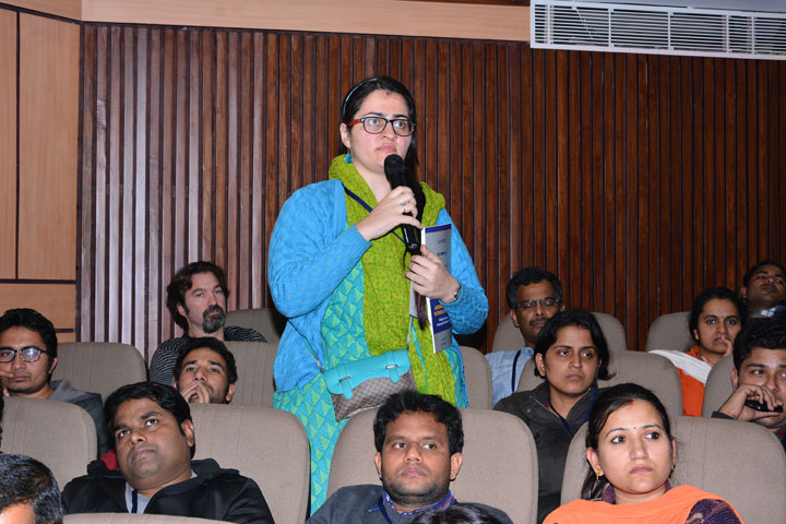
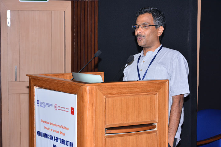
Highly motivated researchers interested in structural biology, particularly, in Cryo- electron microscopy (Cryo- EM) and X-ray crystallography are requested to contact directly to the PI.
Niraj Kumar
Senior Research Fellow
Ankita Arora
Senior Research Fellow
Shivani Sharma
Senior Research Fellow
Soumen Ta
Junior Research Fellow
Tajamul Islam
Integrated MS-PhD student
Ruchika Kumari
Junior Research Fellow (Project)
Tejas Nimkar
Junior Research Fellow (project)
Parthasarathi Behera
Junior Research Fellow (project)
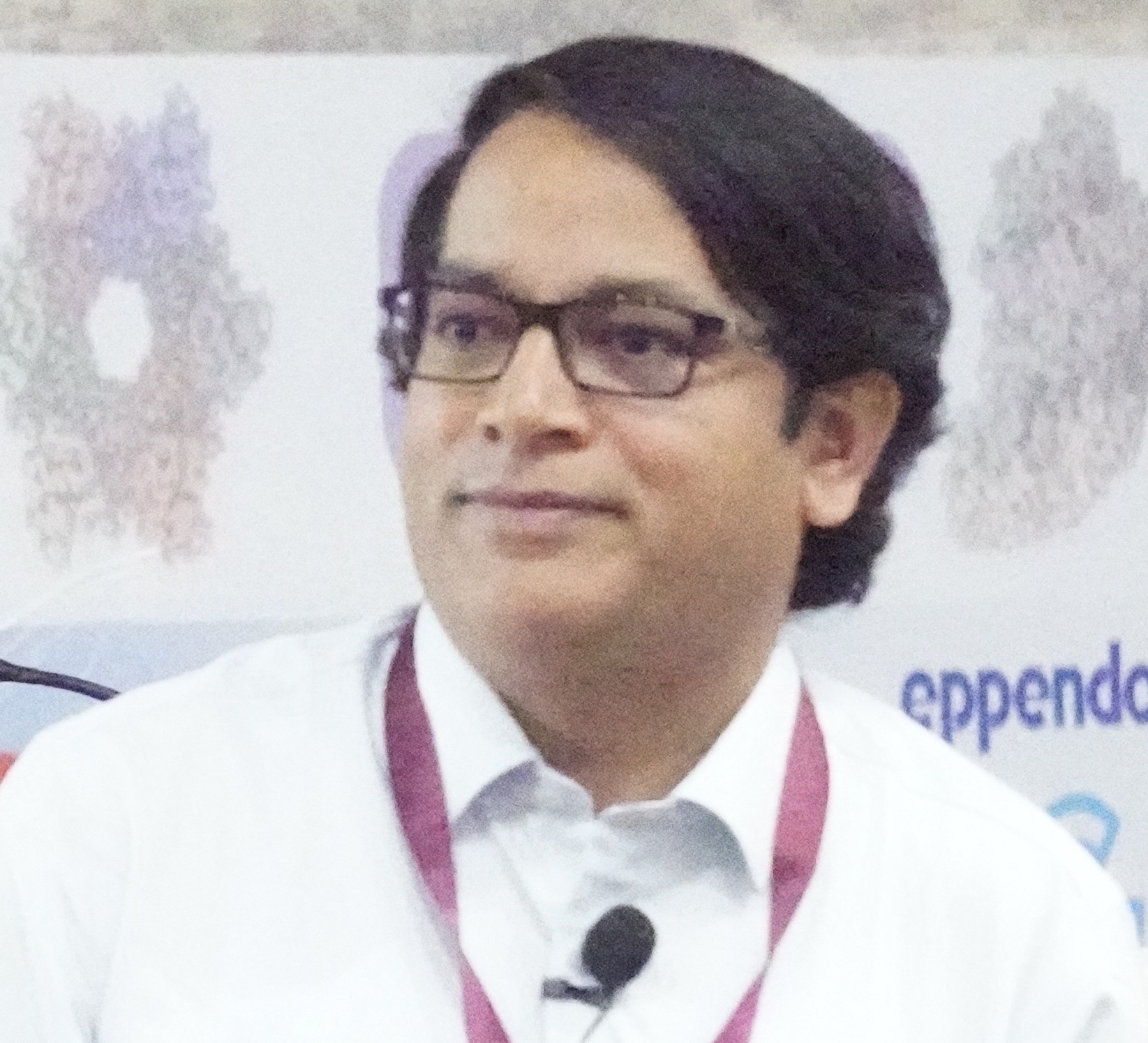
Dr. Prem S. Kaushal
Regional Centre for Biotechnology
NCR Biotech Science Cluster
3rd Milestone, Faridabad-Gurgaon Expressway
P.O. Box No. 3, Faridabad - 121 001
Haryana (NCR Delhi), India
E-mail: prem dot kaushal at rcb dot res dot in
Phone: 91 129-2848980